
Abstract
Cone-beam computed tomography was used to understand the possible correlation between the prevalence of distolingual root (DLR) in permanent mandibular first molars (MFMs) and the associated complicated mandibular incisor’s root canal morphology (MIs) in an Indian population. A total of 400 scans were evaluated for MFMs and MIs. The prevalence of DLRs and root canal anatomy of MIs were assessed based on Vertucci’s classification, and then the sample were grouped according to age, sex and side. Statistical analysis was used to evaluate the possible correlation between the presence of DLRs in the first molar and root canal morphology of incisors. Chi square test was used to evaluate the correlation between the root canal configurations of MIs with the existence of DLRs in MFMs. There was no statistically significant difference between sexes or ages for the prevalence of DLRs in the first molars (p > 0.05), which was 6.62%, with the right side having a greater frequency of DLRs (7.8%) than the left (5.5%). Vertucci Type I canal configuration was most common for the mandibular central (66.75%) and lateral incisors (58.62%). Vertucci Type III was the most common complicated canal morphology, followed by Types V, II, and IV for MIs, with no statistically significant difference in the studied sample’s age and sex. (p < 0.05). No association was observed between the presence of DLRs in first molars and complicated root canal configurations in MIs. Taken together, the possibility of complicated root canal configuration in MIs was lesser in the presence of DLRs in MFMs among the Indian population.
Introduction
Knowledge of tooth and root canal anatomy is crucial for dental practise and for recognising anthropologically significant traits1. Variations in root number and canal morphology are common in permanent mandibular first molars (MFMs), despite the fact that they typically have two roots positioned mesially and distally having three root canals. Many anatomical variations in the roots and root canals of MFMs have been suggested2,3. A major anatomical variation in two-rooted MFMs is the occurrence of an additional root distolingually or mesiobuccally, known as radix entomolaris (RE) or radix paramolaris, respectively, as first reported by Carabelli4.
Speculation surrounds the reasons for of RE formation. Curzon suggested that the “three-rooted molar” characteristic has a high level of genetic penetrance; this dominance was demonstrated by the occurrence of the trait in both pure Eskimo and Eskimo/Caucasian mixes5. According to anatomical studies, some ethnic groups—including populations with Mongoloid features like Chinese, Korean, American Indians, Taiwanese and Eskimo, Korean —have distinct DLRs in their first mandibular molars5,6,7,8,9,10,11,12.
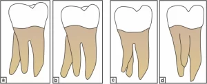
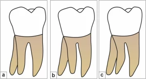
Carlsen and Alexandersen4 (Fig. 1), Ribeiro and Consolaro13 (Fig. 2), and Wang et al.14 have classified RE based on its morphology, buccolingual orientation, and radiographic appearance, respectively. The complexity of the DLR, such as the presence of narrow canals, buccolingual curvature, and short root length, makes biomechanical preparation and obturation of the canal difficult. Moreover, position and superimposition of the DLR over the main distal root may lead to failure in identifying and preparing it, resulting in the failure of endodontic treatment.
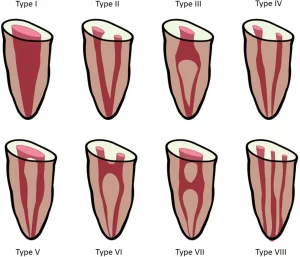
In the literature, there have been reports of variants for each tooth in permanent dentition15. Because it is believed that racial and genetic factors influence root canal configuration, different ethnic populations exhibit varied root canal morphologies16. The most usual morphology of permanent mandibular incisors (MIs) is a single root with a single root canal16 (Fig. 3). However, the root canal morphology of permanent MIs is intricate by the existence of a second canal, lateral canals, and apical deltas, and is not easy to identify17. However, studies have revealed considerable variations in the morphology of the mandibular anterior teeth’s root canal system18,19,20,21. The study conducted by Rankine et al. in the Australian population indicated a high prevalence of two canals in the MIs22, stimulating further research on the canal configuration of other teeth.
The root canal architecture can be studied using ex vivo methods, performed on extracted teeth, or in vivo methods, performed clinically on patients23. The majority of the studies have been conducted in vitro on extracted teeth using invasive techniques such as canal and modified canal staining, tooth-clearing procedures24,25, transverse cross-sectioning26, scanning electron microscopy27, and stereomicroscopy28. Radiography is one of the most essential clinical tools for understanding the internal canal anatomy during endodontic therapy. However, radiography uses two-dimensional (2D) projections of three-dimensional (3D) objects; thus, traditional periapical radiography has certain limitations, such as root distortion and superimposition by surrounding structures29,30,31. Cone-beam computed tomography (CBCT) is regarded as more accurate than conventional radiography and as accurate as the modified canal staining and tooth clearing techniques in detecting root canal systems. CBCT provides 3D images of the teeth and surrounding tissues in three orthogonal planes32. In addition, compared with traditional computed tomography, CBCT delivers a substantially lower effective radiation dosage and is a non-invasive procedure that produces high-quality images.
Although the structural symmetry of root morphology and the root canal configurations between the teeth on both sides of the jaws are of high clinical significance when treating two opposite teeth in the same patient, a study by Wu et al. found a positive correlation in Taiwanese population, also similar findings reported in Cantonese population, but no extensive studies exist on this topic33,34. To the best of our knowledge, information regarding the symmetry and distribution patterns between permanent MFMs with DLRs and permanent MIs with complicated root canal configurations, ipsilaterally and contralaterally among different age groups and sex, using CBCT images has not been reported before in the Indian population.
Therefore, the aim of this study was to observe the prevalence of DLR in the permanent mandibular first molar and complicated canal morphology in permanent MIs in different age groups and sex and to examine whether any association exists between the two in the Indian population using CBCT.
Materials and methods
The research was carried out in accordance with guidelines and regulations set down in the declaration of Helsinki. The current study was approved by the institutional ethics committee of the Nair Hospital Dental College, Mumbai (EC/PG-10/CONS/2019 dated 22/10/2019). The informed consent was waived by the institutional ethics committee of the Nair Hospital Dental College, Mumbai. The CBCT scans were saved in the Digital Imaging and Communications in Medicine (DICOM) format, and these data were saved in an encrypted file confidentially protected yet retrievable if needed.
Sample size calculation
The study sample size was determined based on the expected proportion of findings in the control values estimated from the literature35. Approximately 400 patients/scans were required in the present study, yielding 800 pairs of observations.
Sample selection
CBCT images of the mandibular arch collected from patients who required CBCT scans as part of their treatment were evaluated. The indications for CBCT scanning include the assessment of bone volume for dental implant planning, diagnosis of dentoalveolar trauma, management of impacted teeth before orthodontic treatment, and treatment planning before non-surgical and surgical endodontic treatment.
All of the CBCT scans available from the archives were evaluated retrospectively; based on the inclusion criteria, 400 CBCT scans involving the entire mandibular arch, yielding 800 observations, were included in the study.
Of the 1000 patient CBCT images initially scanned, 400 patients (800 MIs and 800 MFMs) were eligible for further analysis. Of these subjects, 126 (38.5%) were females and 274 (68.5%) were males, with an average age of 44.82 ± 14.89 years (203 samples less than 44 years and 197 above 44 years of age).
Criteria
Inclusion criteria
- (1)Permanent MFMs present bilaterally with complete root formation.
- (2)Permanent mandibular incisors present bilaterally with complete root formation.
- (3)Availability of high-quality CBCT images in which the canal orifice and canal configuration can be easily recognized.
Exclusion criteria
- (1)Endodontically treated teeth.
- (2)Presence of coronal or post-coronal restoration.
- (3)Presence of root resorption/periapical lesions/calcification/open apices.
- (4)CBCT scans having artefacts/poor resolution.
Image acquisition
The evaluated CBCT images were acquired using a CBCT scanner (PLANMECA PROMAX 3D MACHINE) at 90 kV and 10 mA with an exposure time of 27 s. The voxel size of the images was 0.2 mm, and the slice thickness was 0.1 mm. The acquisition process was performed by a licensed radiologist, an expert in operating and acquiring CBCT images according to the manufacturer’s recommended protocol with the minimum exposure necessary for adequate image quality. The “as low as reasonably achievable” protocol was followed.
Image evaluation
The CBCT images were analyzed using Planmeca Romexis imaging software. To ensure optimal visualization, the brightness and contrast of the images were adjusted using the image processing tool embedded in the software.
Two observers, who were endodontists with more than 10 years of experience in the subject, and also experienced in analyzing CBCT images, examined the scans. In order to avoid eye strain, breaks were planned to follow the evaluation of 5 scans, and more than 5 scans were not examined in a row. Inter-examiner bias was resolved by the same observers by analyzing the scans twice within a span of 24 h. The kappa analysis was performed on 10% of total data that is 40 samples before disagreements among examiners were discussed and resolved. There was a near-perfect agreement between Observer 1 and 2 for the presence of Distolingual Root on Right and left side; Vertucci Classification of Central and lateral incisor on the right and left side, (p < 0.05) (Table 1).
The following information was recorded and analyzed:
- 1.The number of roots for permanent mandibular first molar on both sides.
- 2.The root canal configuration [according to Vertucci’s classification FJ16 (Fig. 3) for permanent mandibular central and lateral incisors].
All qualified permanent mandibular incisor and mandibular first molar images were morphologically studied in detail using Planmeca Romexis imaging software.
Mandibular first molar with DLR (RE)
A series of images were assessed from the crown down to the apex in axial sections to identify the presence of DLRs in permanent MFMs.
The scans were further categorized based on the presence or absence of DLRs as follows:
- (1)Non-DLR No DLR was found in the MFMs on either side.
- (2)Unilateral DLR (Uni-DLR) ADLR was found in one mandibular first molar on either the left or right side, and no DLR was found in the other first molar on the contralateral side.
- (3)Bilateral DLR (Bil-DLR) DLRs were found in both the right and left MFMs.
Root canal morphology of permanent MIs
A total of 400 scans were assessed to evaluate the root canal morphology of the permanent mandibular central (n = 800) and lateral (n = 800) incisors on the right and left sides. The root canal configuration of the mandibular central and lateral incisors was determined according to Vertucci’s classification. Several cross-sectional images from the cementoenamel junction to the root apex of the mandibular central and lateral incisors were studied to evaluate the anatomy of the root canals, and the canal configuration was categorized as follows:
- (1)Simple The presence of one root and one canal in the MIs was categorized as a “single canal.”
- (2)Complicated The occurrence of more than one root and canal in the MIs was categorized as a “complicated canal.”
In their respective study on mandibular premolars and mandibular incisors, Liu36 and Wu 35 have categorized Vertucci’s two canal as complicated canal morphology.
The different levels to detect canal morphology was based on the study by Kurumboor K. In this study, the average root length of mandibular central incisor was 12.9 mm and lateral incisor was 12.83 mm. Based on this findings, each section was studied at three levels i.e. 3 mm from apex to study apical third, 6 mm from apex for middle third and 9 mm from apex for coronal third, all three studied in axial section of CBCT scans37.
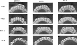
The root canal morphology of the mandibular central and lateral incisors was assessed in sagittal sections at three different levels: 3 mm from the radiographic apex to assess the morphology in the apical third; 6 mm from the apex to assess the morphology of the canals in the middle third of the root; and 9 mm from the apex to assess the morphology of the canals in the coronal third of the root.
The canal configuration and the numbers of the roots and canals of the central and lateral incisors were classified according to Vertucci’s classification16.
Statistical analysis
The data was obtained and entered in Microsoft Excel Version 13. Using IBM SPSS Version 21, statistical analysis of the data was performed. The Data was observed to be categorical for multiple variables hence frequency and proportion was obtained for continuous variable like age; Mean and SD was obtained. Descriptive statistics were expressed as means and standard deviations, frequencies, or percentages, as appropriate, of each measurement calculated at the subject and/or tooth levels. The chi-square test was used for examining differences in categorical variables such as side (left vs right), age (44 ± 52 years) and gender (male vs female) with DLR group (Non-DLR, Uni-DLR, or Bil-DLR). To compare between Gender and Vertucci Classification and Age Categorization and Vertucci Classification, Mann Whitney U test was applied. The association between the prevalence of DLRs and the complex root canal morphology of incisors were analyzed using the chi-square test and SPSS 17.0 software (SPSS Inc, Chicago, IL). All the statistical tests were applied keeping confidence interval at 95% and (p < 0.05) was considered to be statistically significant.
Results
Prevalence of DLRs
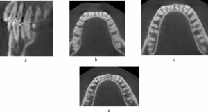
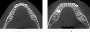
A total of 400 scans exhibiting 800 MFMs were assessed for the presence of DLR. Of the 800 MFMs evaluated, 6.62% (53 of 800) exhibited DLR. Of the total 800 MFMs evaluated, 7.8% (31 out of 400) right and 5.5% (22 out of 400) left MFMs exhibited distolingual roots (p > 0.01)(Table 2) (Fig. 4). Of the total 400 scans examined, 3.5% (14 out of 400) showed the unilateral presence of DLRs. However, the prevalence of bilateral DLRs in the MFMs was found to be 5% (20 out of 400) (Fig. 5).According to the gender-wise distribution of DLR, 9.5% (38 out of 400) males and 3.75% (15 out of 400) females showed presence of DLR (p > 0.05 on right and left) (Table 3). Age-wise distribution of DLR in MFM showed more prevalence in the age group above 44 years on both the right and left side that is 8.5% (34 out of 400) and 4.75% (19 out of 400) in groups less than 44 years (p > 0.05 on right and left) (Table 4). There was no statistically significant difference in the distribution of DLR in MFM among age, sex, and side (p > 0.05).
Mandibular central incisors
Overall, 66.75% (534 of 800) of the mandibular central incisors showed a simple (Vertucci Type I) root canal morphology. Of the 800 central incisors evaluated, 68% (272 of 400) on the right and 65.5% (262 of 400) on the left side showed a simple canal morphology (Table 5) (p > 0.01).
Approximately 33.25% (266 of 800) of the mandibular central incisors showed a complicated root canal morphology (Type III, V, or II). Complicated root canal morphology including Vertucci’s Type II, III, IV, and V was found in 32% (128 of 400) of MCIs on the right side and 34.5% (138 of 400) of the left side. Vertucci Type III (28.3% on the right side and 31% on the left side) canal configuration was the second most common root canal morphology for the mandibular central incisors, followed by Type V and II (p > 0.01) (Table 5).
MCIs, on both the right and left side showed Vertucci’s Type I morphology more commonly in males (66.8–68.6%) than females (62.7–66.8%) (p > 0.05 on right and left) (Table 6). Also, the prevalence of complicated root canal morphology of MCIs was more in males than females. Age wise distribution of root canal morphology shows more prevalence of Type I root canal configuration in age groups below 44 years and more prevalence of complicated root canal morphology in age above 44 years on both right and left sides. (p > 0.05 on right and left).
Regarding age, gender, and side, there was no statistically significant variation in the distribution of root canal morphology in MCIs (p > 0.05).
Mandibular lateral incisors
Overall, 58.62% (469 of 800) of the mandibular lateral incisors showed a simple (Vertucci Type I) root canal morphology. Of the 800lateral incisors evaluated, 58.3% (233 of 400) on the right and 59% (236 of 400) on the left side showed a simple canal morphology (p > 0.01) (Table 5) (Fig. 6).
Approximately 41.37% (331of 800) of the mandibular lateral incisors showed a complicated root canal morphology (Type III, V, or II). Complicated root canal morphology including Vertucci’s Type II, III, IV, and V was found in 41.75% (167 of 400) of MCIs on the right side and 41% (164 of 400) of the left side. Vertucci Type III (34.8% on the right side and 35.8% on the left side) canal configuration was the second most common root canal morphology for the mandibular lateral incisors, followed by Type V and II (p > 0.01) (Table 5).
MLIs, on both the right and left side showed Vertucci’s Type I morphology more commonly in males (59–59.9%) than females (56.3–57.1%) (Table 6). Also, the prevalence of complicated root canal morphology of MCIs was more in males than females (p > 0.05 on right and left). Age wise distribution of root canal morphology shows more prevalence of Type I root canal configuration in age groups below 44 years and more prevalence of complicated root canal morphology in age above 44 years on both right and left sides (p > 0.05 on right and left) (Table 7).
Regarding age, gender, and side, there was no statistically significant variation in the distribution of root canal morphology in MCIs (p > 0.05).
Association Between DLR and the Root Canal Morphology of Permanent MIs
In relation to the central incisors
Overall, the prevalence of complicated root canal configuration of the mandibular right and left central incisors in the presence of the first molar with DLR was 32% (128 of 400) and 34.5% (138 of 400) respectively. Approximately 7.8% (31 of 400) and 5.5% (22 of 400) of the permanent MFMs exhibited DLRs on the right and left sides, respectively. In the presence of DLRs, 27.2% (9 of 33) and 30.3% (10 of 33) of the central incisors showed complicated root canal configurations on the right and left sides, respectively. In the absence of DLRs, 32.4% (119 of 367) and 34.8% (128 of 367) of the central incisors showed complicated root canal morphology on the right and left sides, respectively. Therefore, the frequency of complicated root canal morphology in MCIs was more in the absence of DLR in MFMs as compared to the presence of DLR (p > 0.05 on right and left).
Read more >>https://www.nature.com/articles/s41598-024-51198-1